Wenn Sie suchen nach Former NFL player confirmed as 1st diagnosis of CTE in living patient Du hast besuchte Nach rechts Webseite. Wir haben 35 Bilder etwa Former NFL player confirmed as 1st diagnosis of CTE in living patient wie MRI front view Stock Photo – Alamy, Could Structural MRI Findings Help Detect CTE During Life? und auch Preoperative MRI T1 with contrast, CTA, and ophthalmic examination. Hier ist es:
Former NFL Player Confirmed As 1st Diagnosis Of CTE In Living Patient
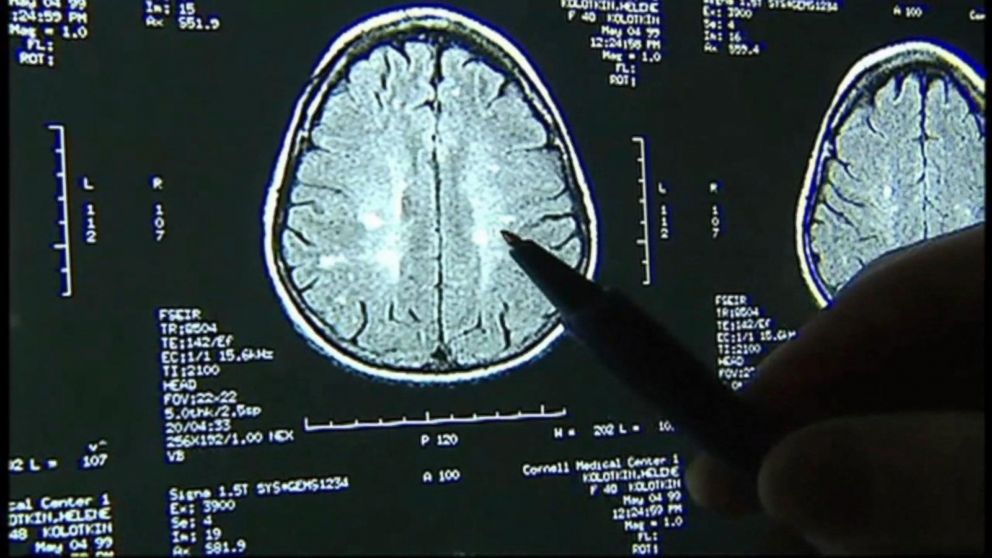
abcnews.go.com
cte khou mild
Pre-operative MRI And CECT. A-C: Axial Views On MRI. A: T1-weighted

www.researchgate.net
Imaging May Predict CTE – International College Of Chiropractic

chironeurointl.org
My MRI Scan : R/oddlyterrifying

www.reddit.com
MRI T2 TSE In Coronal (a) And Transverse (b) Views Show The Neck Of The

www.researchgate.net
Image Movement Due To Tube CTE – Experienced Deep Sky Imaging – Cloudy

www.cloudynights.com
MRI T2 TSE In Coronal (a) And Transverse (b) Views Show The Neck Of The

www.researchgate.net
Premium Vector | Mass Rapid Transit Train Front View

www.freepik.com
Premium Photo | MRI Or CT Scan Medical Diagnosis Machine Background Ai

www.freepik.com
Setting Up An MRI Protocol | Perry Lab For Craniofacial Imaging | ECU

cahs.ecu.edu
-Preoperative MRI: Axial TSE-T1 View After Administration Of Contrast

www.researchgate.net
Room With A White Mri Machine Background, Picture Of Cat Scan Machine

pngtree.com
MRI Images In Axial Plan In T1 Weighted FSE (a), T2 Weighted FSE (b

www.researchgate.net
Magnetic Resonance (MRI) Coronal Image TSE -T1-weighted After

www.researchgate.net
Comparison Of The (a) MSCT Images, (b) Corresponding ZTE-MRI And (c

www.researchgate.net
MRI Front View Stock Photo – Alamy

www.alamy.com
mri front alamy
Preoperative MRI T1 With Contrast, CTA, And Ophthalmic Examination

www.researchgate.net
Control Brain MRI, In Axial Sections, T1 WI (A) And T1 POST Contrast

www.researchgate.net
MRI Scans May Be Useful In Diagnosing Chronic Traumatic Encephalopathy

neurosciencenews.com
mri neurosciencenews cte brain ucla encephalopathy scans traumatic chronic diagnosing useful may researchers regions shrinkage consistent several key technology found
CHAPTER-15

www.cis.rit.edu
mri nex tr te matrix ms
A,B) Axial T2-weighted TSE MRI. Coronal T2-weighted TSE MRI (C) And

www.researchgate.net
Could Structural MRI Findings Help Detect CTE During Life?
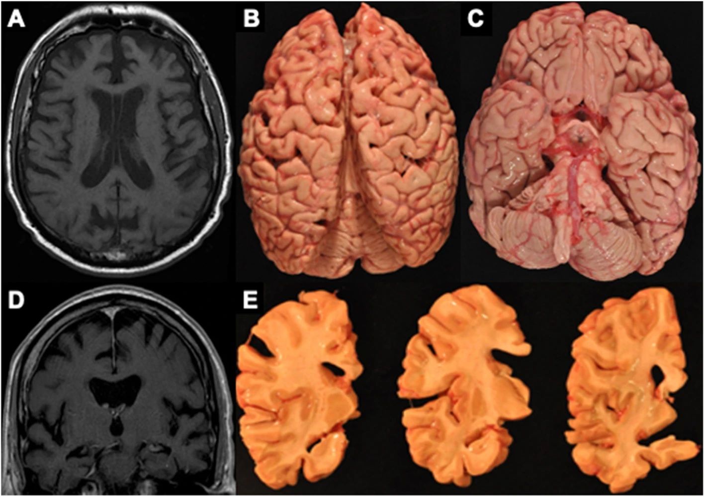
www.diagnosticimaging.com
MRI And Contrast-enhanced Computed Tomography (CECT) Images At The

www.researchgate.net
Development Of Zero TE MRI To Visualise Cranial Bones, Nerves And

www.imagingcdt.com
Front View Of The FFC-MRI Scanner Presented Here. The Scanner Has A

www.researchgate.net
Are There Early Indicators Of CTE In The Brains Of Epilepsy Patients
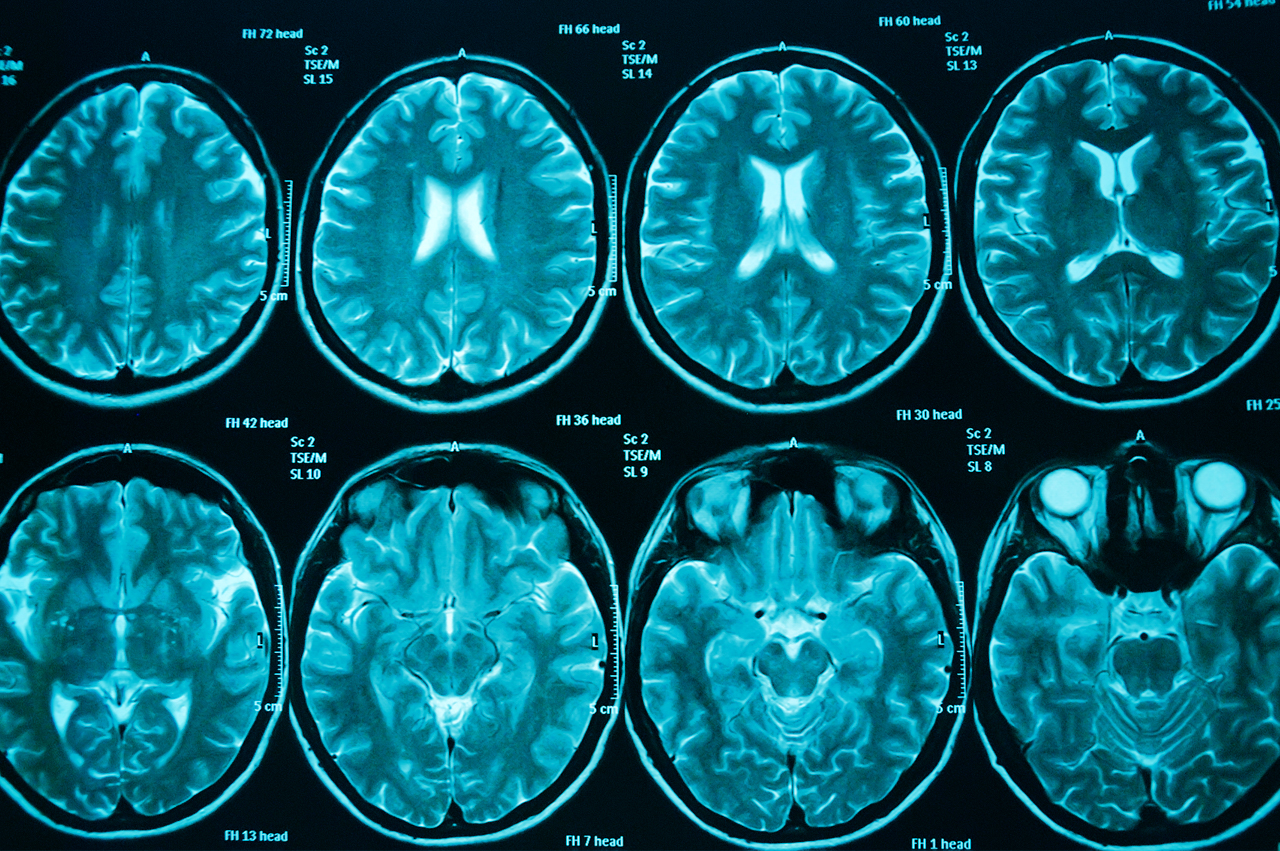
www.neuro-central.com
scans brain cte tumour neuro ward diagnosis brains neurosurgery life day after patients epilepsy central ot treatment diagnostic
Premium Photo | Modern MRI Machine In A Room

www.freepik.com
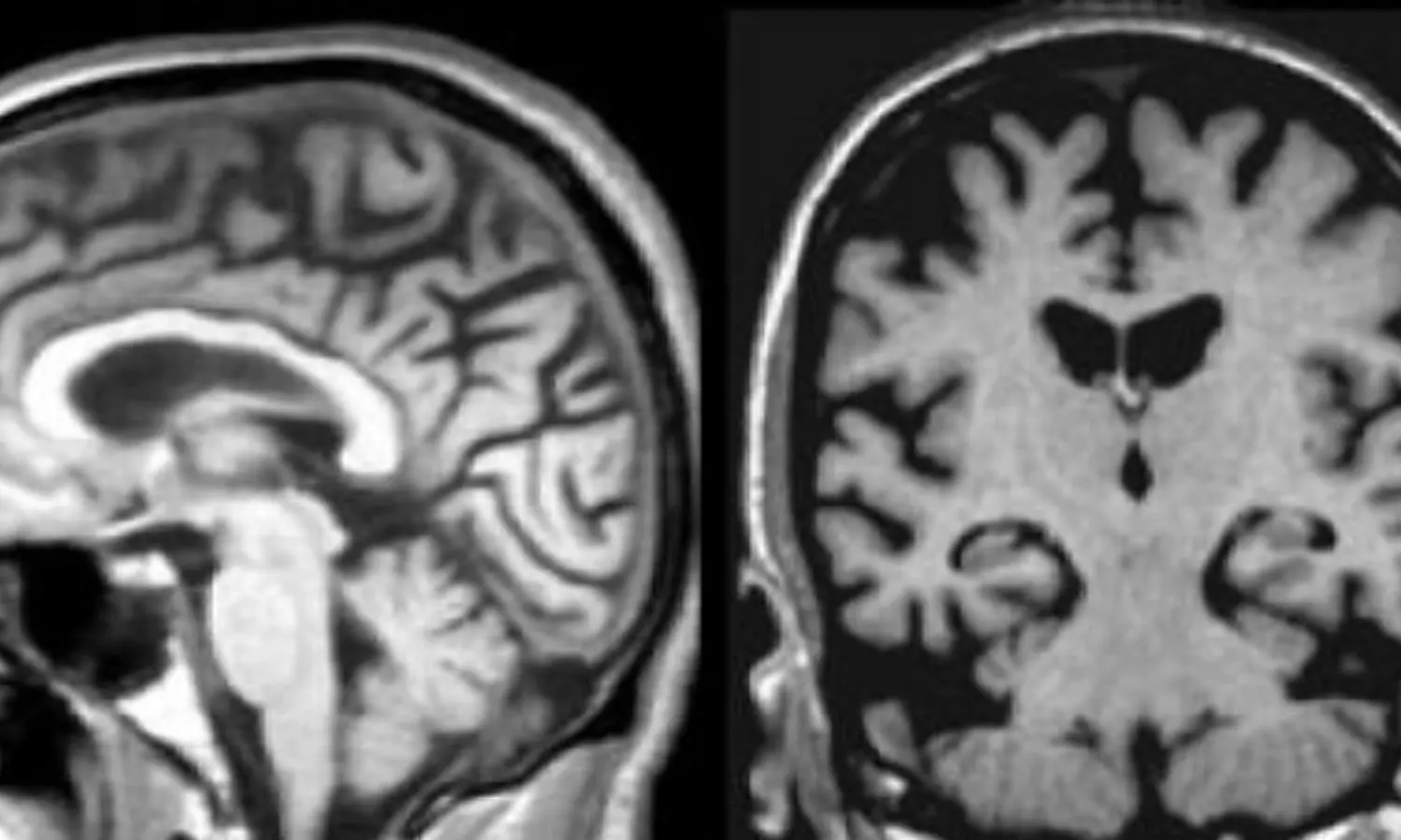
MRI May Detect Concussion-Linked CTE While Patients Are Still Alive
medicaldialogues.in
MRI Reveals Changes Related To Possible CTE. Axial Image Reveals Both

www.researchgate.net
CHAPTER-15

www.cis.rit.edu
mri ms pathology te tr nex matrix head chap
Axial Views For One Patient Of The MRI, The CT And The Pseudo-CTs

www.researchgate.net
CHAPTER-15

www.cis.rit.edu
mri neck head side sagittal ms spot gif edu what te nex tr matrix rit cis chap
Preoperative MRI Images. (a) Sagittal T1-weighted Contrast-enhanced

www.researchgate.net
Coronal MRI Section Showing T1 Image Contrast‐enhanced | Download

www.researchgate.net
First MRI. The Next Day, After CT Scan Was Preformed, Axial T2W_TSE 5

www.researchgate.net
Premium photo. Development of zero te mri to visualise cranial bones, nerves and. First mri. the next day, after ct scan was preformed, axial t2w_tse 5

